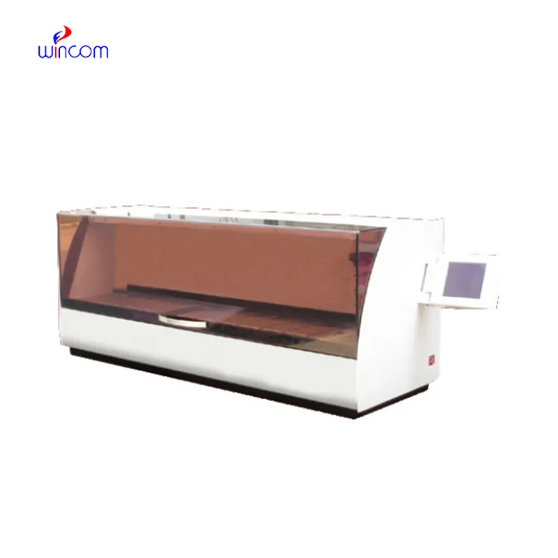
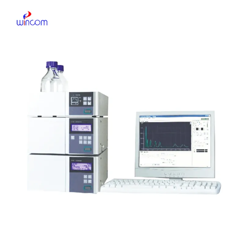
preparative hplc system offers high resolution separation of complex samples in clinical, pharmaceutical, and hospital laboratories, thereby supporting advanced laboratory workflows. It allows performing an in-depth analysis of drugs, metabolites, and small biomolecules. preparative hplc system is used by laboratory staff for research validation, patient monitoring, and method development. Its precision, speed, and adaptability make analytical efficiency greater and at the same time, make consistent and reproducible results which in turn, strengthen laboratory operations in the areas of healthcare and scientific environments.
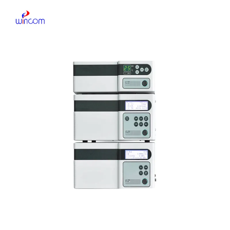
preparative hplc system finds use in clinical toxicology laboratories to pinpoint and measure the amounts of possible poisons or drugs in abuse samples taken from patients. It is based on the separation of the various substances from complex mixtures like blood or urine, and that information is very important for the hospital doctors, who will then diagnose the case, decide on the treatment and monitor the patient’s safety.
Advanced software platforms for predictive analytics in healthcare are going to be part of the preparative hplc system integration. The hospitals will take advantage of the real-time data provided by the patient samples to influence their clinical decisions. Molecular profiling as well as automated quality control and laboratory efficiency will be thepreparative hplc system future applications targeting the improvement of patient care.
Routine upkeep of preparative hplc system is of utmost importance in clinical laboratories to maintain the accuracy of patient sample analysis. Regular cleaning of pipes, changing of deteriorated seals and calibration of measuring instruments will block adulteration and keep the latter's sensitivity. Lab personnel must record maintenance activities and keep watch over system performance. Constant attention guarantees that preparative hplc system provides dependable, reproducible results for hospital diagnosis and research work.
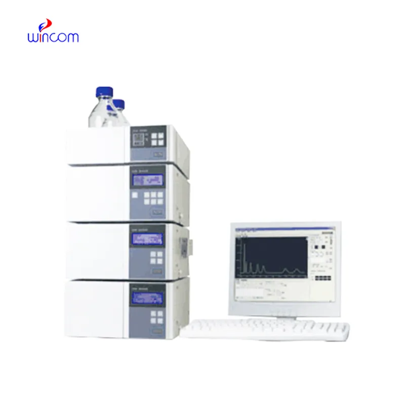
preparative hplc system are a major factor in the daily activities of pharmaceutical labs, as they are used for verifying drug formulations, detecting impurities, and making sure that quality standards are met. It provides accurate quantification by separating active ingredients from excipients. Lab scientists utilize this for process optimization and stability evaluation under varied conditions. By providing reproducible analytical data, preparative hplc system assists in both method validation and research development. Its accuracy guarantees that pharmaceutical products will be compliant with regulations. In lab environments, preparative hplc system is a time-saving method not only for compound profiling but also for comprehensive analyses, thus being a fundamental tool in the quality control of pharma and research labs dealing with drug development.
Q: Do you need special training for HPLC operation? A: The answer is yes, training is a prerequisite to accurately and safely using pumps, columns, and detectors. Q: What type of maintenance does HPLC have? A: It requires cleaning, flushing, and inspection of all components as well as calibrating. Q: Is it possible to use HPLC in drug monitoring? A: Sure, it is a common practice in hospitals to monitor the levels of therapeutic drugs and also to identify metabolites in the samples taken from the patients. Q: What is the duration of analysis using HPLC in a typical case? A: The analysis time can range from a few minutes to more than an hour depending on the nature of the sample and the kind of column used. Q: Is HPLC a good choice for environmental testing? A: Yes, it can be used to find out the presence of pollutants, pesticides, and other harmful substances in water, soil, and air samples.
The microscope delivers incredibly sharp images and precise focusing. It’s perfect for both professional lab work and educational use.
This x-ray machine is reliable and easy to operate. Our technicians appreciate how quickly it processes scans, saving valuable time during busy patient hours.
To protect the privacy of our buyers, only public service email domains like Gmail, Yahoo, and MSN will be displayed. Additionally, only a limited portion of the inquiry content will be shown.
Hello, I’m interested in your centrifuge models for laboratory use. Could you please send me more ...
Could you please provide more information about your microscope range? I’d like to know the magnif...
E-mail: [email protected]
Tel: +86-731-84176622
+86-731-84136655
Address: Rm.1507,Xinsancheng Plaza. No.58, Renmin Road(E),Changsha,Hunan,China

 af
af
 es
es
 ar
ar
 tr
tr
 sw
sw
 pt
pt
 th
th
 ur
ur
 bn
bn
 ne
ne
 vi
vi
 km
km
 lo
lo
 de
de
 ru
ru
 fi
fi
 nl
nl
 fa
fa
 fr
fr
 ko
ko