The pocket ultrasound scanner works by integrating cutting-edge image optimization features that reduce artifacts and improve detail recognition thereby increasing diagnostic accuracy. It caters to multilingual users and sets for personal usability requirements for global needs. The device provides imaging of the same quality regardless of the patient type or clinical condition.

The pocket ultrasound scanner has a very wide use in radiology where it supports the guidance of non-invasive procedures. It is very important in gynecology where it is allowed to conduct reproductive system evaluations. In orthopedics, the pocket ultrasound scanner helps visualize muscles, joints, and tendons ensuring correct diagnostic interpretation. It is this very nature of versatility that makes it a must-have for imaging during procedures in real time.

The pocket ultrasound scanner can look forward to getting advantages from miniaturization and wearable technologies. Portable or handheld versions of the pocket ultrasound scanner will become more widespread to facilitate quick diagnoses in rural as well as emergency setups. The integration of telemedicine services will thus facilitate concurrent consultations via the pocket ultrasound scanner.

The pocket ultrasound scanner needs to be maintained based on the manufacturer's recommendations. The transducers should be kept in specialized holders. The cleaning agent should be non-corrosive. The electrical contacts should remain dry. Functional tests should be carried out on a regular basis to ensure that the pocket ultrasound scanner functions properly and remains a safe instrument.
Built for performance and accuracy, the pocket ultrasound scanner is an imaging diagnosis platform. It gives real-time images of tissues, organs, and vascular systems, enabling increased detection of pathology. Space-efficient and compact, the pocket ultrasound scanner is ideal for hospitals, clinics, and ambulatory healthcare facilities. Its accuracy imaging enables physicians to offer timely and informed medical care.
Q: What imaging modes are available on the ultrasound scannert? A: It supports multiple modes such as B-mode, M-mode, and color Doppler for diverse diagnostic applications. Q: How does the ultrasound scannert improve diagnostic accuracy? A: By providing high-resolution images and real-time feedback, it enables more precise medical evaluations. Q: Can the ultrasound scannert be used in field or remote settings? A: Yes, its portable versions are designed for mobility and can be used in clinics, hospitals, or mobile healthcare units. Q: What kind of display does the ultrasound scannert use? A: It typically features a high-definition digital display that enhances image visualization and readability. Q: How is data from the ultrasound scannert managed? A: The device allows secure storage, easy access, and export of imaging data through USB or network connections.
The delivery bed is well-designed and reliable. Our staff finds it simple to operate, and patients feel comfortable using it.
We’ve been using this mri machine for several months, and the image clarity is excellent. It’s reliable and easy for our team to operate.
To protect the privacy of our buyers, only public service email domains like Gmail, Yahoo, and MSN will be displayed. Additionally, only a limited portion of the inquiry content will be shown.
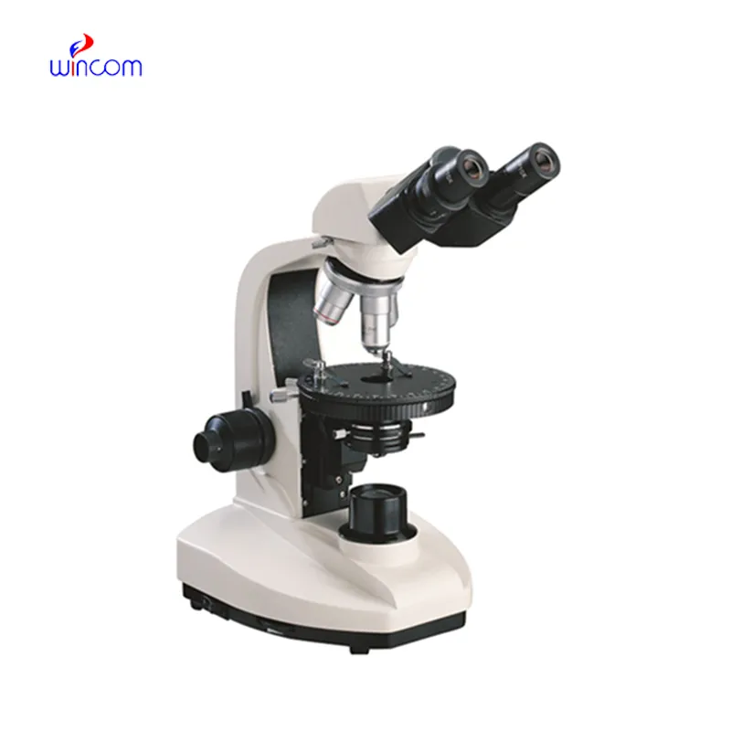
Could you please provide more information about your microscope range? I’d like to know the magnif...
Could you share the specifications and price for your hospital bed models? We’re looking for adjus...
E-mail: [email protected]
Tel: +86-731-84176622
+86-731-84136655
Address: Rm.1507,Xinsancheng Plaza. No.58, Renmin Road(E),Changsha,Hunan,China

 af
af
 es
es
 ar
ar
 tr
tr
 sw
sw
 pt
pt
 th
th
 ur
ur
 bn
bn
 ne
ne
 vi
vi
 km
km
 lo
lo
 de
de
 ru
ru
 fi
fi
 nl
nl
 fa
fa
 fr
fr
 ko
ko