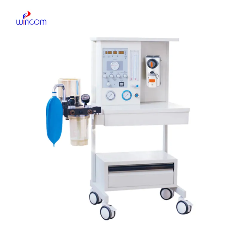
mri compatible anesthesia machine has been specifically designed to administer and manage anesthesia in a controlled manner while also providing the necessary mechanical ventilation and the monitoring of vital signs, which is done continuously. It brings together the monitoring of oxygen, the measurement of airway pressure, and the tracking of respiration, hence clinicians can modify the amount of sedation quickly and easily. This system is adopted by hospitals and research labs to guarantee patient safety during the times of operation and experiments. The integration of safety alarms, vaporizers, and variable gas flow ensures performance that is precise and reliable. mri compatible anesthesia machine offers better safety during procedures, easier and more effective anesthesia management, and importantly, it leaves the door open for high-quality patient care through physiological feedback.
mri compatible anesthesia machine is being practiced in emergency departments as a means of fast anesthesia induction during urgent surgical interventions. Trauma cases usually have unstable patients, which makes precise management of ventilation and anesthetic delivery very important. The apparatus gives the medical personnel the opportunity to rapidly adapt gas flow and airway pressure to the patient's reaction. The introduction of the device is facilitating the management of the airway securely during the period of emergency intubation and immediate operative care. By its dependable function in emergencies, mri compatible anesthesia machine not only helps but also hospitals to provide anesthesia that is effective during emergencies that are critically timed.
Future advancements in mri compatible anesthesia machine might have better user interfaces that can be user-friendly and save the time for training. Visual aids that are easy to understand and controls that are user-friendly can make it possible to have the same quality of use across different clinical teams. Those hospitals that constantly have new staff coming in and out or are using different medical specialists may find the new features of mri compatible anesthesia machine very helpful in dealing with the problems caused by the diversity of the staff. In focusing on the characteristics of the product that make it easier to use and more effective, mri compatible anesthesia machine will be able to provide support for the safe management of anesthesia even in situations where the healthcare workforce is changing.
mri compatible anesthesia machine are the most important things in hospital maintenance and cleaning procedures. The external surfaces need to be disinfected with approved disinfectants to totally avoid the transfer of germs. For example, the internal parts like vaporizers need to be checked regularly to make sure that the residue does not harm the operation. In the labs and operating rooms, highly strict cleanliness rules are applied which are beneficial to both patients and medical personnel. Moreover, proper drying after cleaning saves the equipment from moisture damage. These practices of care and maintenance ensure the safe use and keep the mri compatible anesthesia machine constant in all the medical departments performance.
Hospital intensive care units make use of mri compatible anesthesia machine for sedation and ventilation support in critically ill patients. The instrument guarantees the patient's safety during the surgical interventions because it is capable of delivering precise anesthetics and monitoring the patient's respiration at the same time. It has alarm systems and customizable settings that enable quick reaction to changes in the body's functions. By ensuring dependable anesthesia and ventilation support, mri compatible anesthesia machine becomes a crucial factor in the provision of safe and high-quality patient care in the areas of surgery and critical care labs.
Q: How frequently should Anesthesia Machine be checked? A: The machine should be routine checked before daily use and also during the scheduled maintenance cycles. Q: Is the Anesthesia Machine movable within a hospital? A: There are lots of models available that are specifically designed with wheels that allow easy movement between the departments. Q: Can it be connected with hospital monitoring systems? A: There are some models that are designed for the connection with central monitoring and data systems. Q: What is the procedure when the oxygen supply is cut off? A: The safety mechanisms will alert the clinicians and will also help in keeping the patient protected. Q: Is the use of this equipment limited to specially trained personnel only? A: Definitely, only properly trained personnel will be able to use the machine safely and efficiently.
The centrifuge operates quietly and efficiently. It’s compact but surprisingly powerful, making it perfect for daily lab use.
I’ve used several microscopes before, but this one stands out for its sturdy design and smooth magnification control.
To protect the privacy of our buyers, only public service email domains like Gmail, Yahoo, and MSN will be displayed. Additionally, only a limited portion of the inquiry content will be shown.
We’re interested in your delivery bed for our maternity department. Please send detailed specifica...
We’re looking for a reliable centrifuge for clinical testing. Can you share the technical specific...
E-mail: [email protected]
Tel: +86-731-84176622
+86-731-84136655
Address: Rm.1507,Xinsancheng Plaza. No.58, Renmin Road(E),Changsha,Hunan,China

 af
af
 es
es
 ar
ar
 tr
tr
 sw
sw
 pt
pt
 th
th
 ur
ur
 bn
bn
 ne
ne
 vi
vi
 km
km
 lo
lo
 de
de
 ru
ru
 fi
fi
 nl
nl
 fa
fa
 fr
fr
 ko
ko