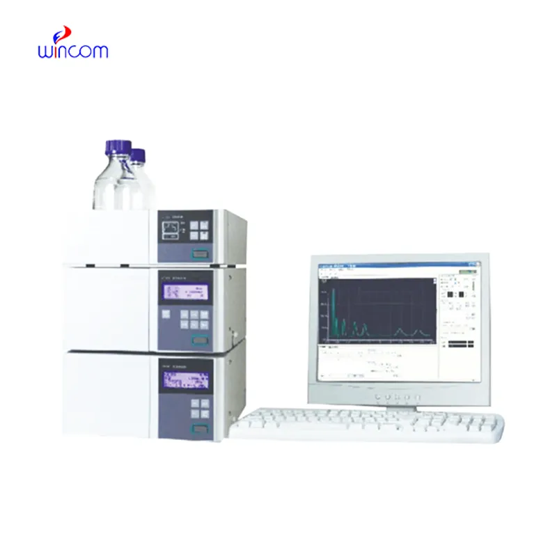
monolithic hplc column is a primary tool in hospital and laboratory analytics. Its skills of isolating, measuring, and characterizing both chemical and biological substances enhance research as well as clinical testing. Quality control, drug testing, and testing of samples are done by laboratory technicians using monolithic hplc column. The device's flexibility and reliability guarantee uniform performance, yielding critical analytical data that are vital for patient care, experimental validation, and smooth and fast laboratory operations in both healthcare and scientific domains.
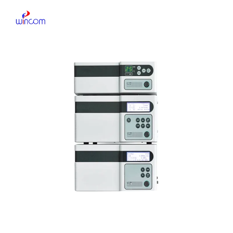
monolithic hplc column are utilized by clinical laboratories for hormone and endocrine-related biomarker detection. It delivers trustworthy information for the diagnosis of endocrine diseases by correctly separating substances like cortisol, thyroid hormones, or insulin. Techs in a laboratory rely on monolithic hplc column to give accurate and repeatable results, thereby helping doctors in individual treatment plan.
Advanced software platforms for predictive analytics in healthcare are going to be part of the monolithic hplc column integration. The hospitals will take advantage of the real-time data provided by the patient samples to influence their clinical decisions. Molecular profiling as well as automated quality control and laboratory efficiency will be themonolithic hplc column future applications targeting the improvement of patient care.
Systematic cleaning, pressure monitoring, and timely worn parts replacement are among the measures to be taken in the hospital laboratories to keep monolithic hplc column under control. Laboratory staff must ensure the observance of the suggested operating conditions, avoid the formation of air bubbles in the system, and check for proper solvent compatibility. Regular maintenance maintains the performance of the column, avoids contamination, and allows the analysis to be precise and reproducible, thereby benefiting not only routine patient testing but also experimental research.
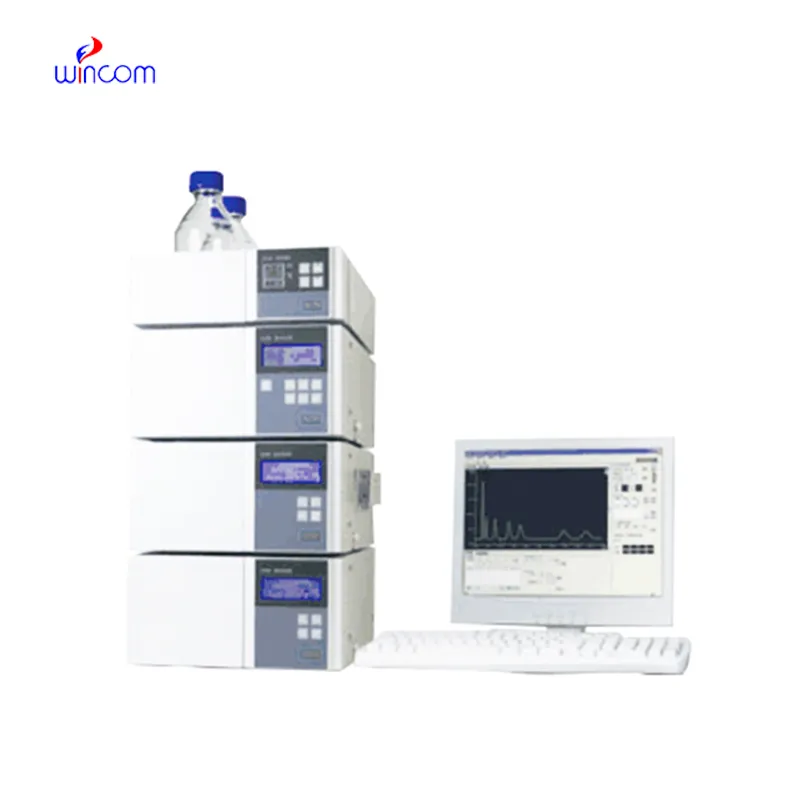
monolithic hplc column is of utmost importance in biochemistry laboratories of both universities and hospitals. It makes detailed study of proteins, peptides, and metabolites possible through the separation of intricate mixtures. The application of it includes but is not limited to enzymatic analysis, biomarker detection, and data obtained through metabolomics. The sensitivity and reproducibility of the device guarantee genuine molecular profiles. Lab technicians make use of monolithic hplc column to conclude their experiments and provide evidence for scientific publications. Its accuracy and versatility give biochemistry labs the ability to perform cutting-edge research in molecular mechanisms, disease pathways, and therapy targets thus, it becomes an indispensable tool for both analytical and clinical lab investigations.
Q: What is the sample preparation for HPLC? A: For the most part, samples should be filtered, diluted, or subjected to solvent extraction in order to avoid column clogs and have the results be accurate Q: Is HPLC able to pick trace-level compounds? A: With the right detectors, it can pick up such substances in extremely small amounts with high sensitivity. Q: Is HPLC a method that can be applied to analysis of proteins? A: Yes, particularly if one employs size-exclusion and reversed-phase columns for protein, peptide, and biomolecule separation. Q: What is the process of calibrating HPLC? A: The process is done by taking standards of known concentrations that are the same as the one in the sample and using them to check the performance of the column and the accuracy of the detector. Q: Are particular solvents needed for HPLC? A: Yes, the solvents used need to be compatible with the type of the column and the detectors to prevent any damage or interference in the analysis process.
The delivery bed is well-designed and reliable. Our staff finds it simple to operate, and patients feel comfortable using it.
This ultrasound scanner has truly improved our workflow. The image resolution and portability make it a great addition to our clinic.
To protect the privacy of our buyers, only public service email domains like Gmail, Yahoo, and MSN will be displayed. Additionally, only a limited portion of the inquiry content will be shown.
Hello, I’m interested in your centrifuge models for laboratory use. Could you please send me more ...
We are planning to upgrade our imaging department and would like more information on your mri machin...
E-mail: [email protected]
Tel: +86-731-84176622
+86-731-84136655
Address: Rm.1507,Xinsancheng Plaza. No.58, Renmin Road(E),Changsha,Hunan,China

 af
af
 es
es
 ar
ar
 tr
tr
 sw
sw
 pt
pt
 th
th
 ur
ur
 bn
bn
 ne
ne
 vi
vi
 km
km
 lo
lo
 de
de
 ru
ru
 fi
fi
 nl
nl
 fa
fa
 fr
fr
 ko
ko