Today, clinical laboratories always rely on hplc-ms for the purpose of giving comprehensive chemical and biological data from patient samples. The technology's exceptional sensitivity and accuracy make it possible to separate even the smallest amounts of substances such as drugs and metabolites from complicated mixtures. Laboratory staff performs using hplc-ms in method development, validation and ongoing monitoring of the lab's analytical performance. The multi-use of the instrument guarantees its presence during both normal testing and research work, hence hospitals and laboratories are always consistent in providing accurate and trustworthy diagnostic and analytical results.
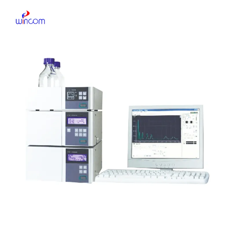
hplc-ms finds extensive application in hospital laboratories for monitoring drugs therapeutically. It provides precise determination of drug levels in patients' samples, thus making safe and effective dosing possible. Metabolites are tracked, treatment progress is assessed, and unexpected drug interactions are detected by the laboratory personnel. Its high accuracy and reproducibility facilitate both medical decision-making and research, hence, hplc-ms becomes an indispensable instrument in taking care of patients and analyzing the medical field.

hplc-ms is assigned to become an important player in translational research which is being conducted in hospitals. Among the future developments are the combined detection systems, quicker analysis cycles, and improved reproducibility. hplc-ms will be the mainstay of hospitals' molecular profiling and drug testing along with patient monitoring thus facilitating hospital diagnostics and personalized medicine research.
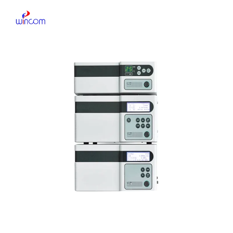
The effectiveness of a laboratory is determined by the proper maintenance of hplc-ms. If the pump seals are regularly cleaned, the flow rates are monitored, and the usage of incompatible solvents is avoided then damage to the laboratory equipment can be prevented. It is essential for the technicians to carefully examine the columns, detectors, and tubing and in case of any sign of wear to conduct the scheduled calibration. Keeping hplc-ms in their best condition guarantees reproducibility, lowers the risk of equipment breakdown, and provides continuous performance for both hospital tests and experiments.
hplc-ms is of utmost importance in biochemistry laboratories of both universities and hospitals. It makes detailed study of proteins, peptides, and metabolites possible through the separation of intricate mixtures. The application of it includes but is not limited to enzymatic analysis, biomarker detection, and data obtained through metabolomics. The sensitivity and reproducibility of the device guarantee genuine molecular profiles. Lab technicians make use of hplc-ms to conclude their experiments and provide evidence for scientific publications. Its accuracy and versatility give biochemistry labs the ability to perform cutting-edge research in molecular mechanisms, disease pathways, and therapy targets thus, it becomes an indispensable tool for both analytical and clinical lab investigations.
Q: What types of HPLC columns are available? A: Reversed-phase, normal-phase, ion-exchange, and size-exclusion columns are the main types of columns used according to the nature of the analytes. Q: Can multiple samples be analyzed simultaneously? A: Yes, in high-throughput systems, automated sample injection and sequential analysis are among the techniques to achieve this. Q: How does temperature affect HPLC performance? A: Temperature changes can cause variations in separation efficiency and retention times; however, the majority of labs make use of precise temperature control. Q: Can HPLC be integrated with data software? A: Sure, it can be linked with laboratory software for data collection, processing, and reporting. Q: What types of laboratories use HPLC? A: HPLC is employed by hospitals, pharmaceuticals, biochemistry research, and environmental testing labs.
The centrifuge operates quietly and efficiently. It’s compact but surprisingly powerful, making it perfect for daily lab use.
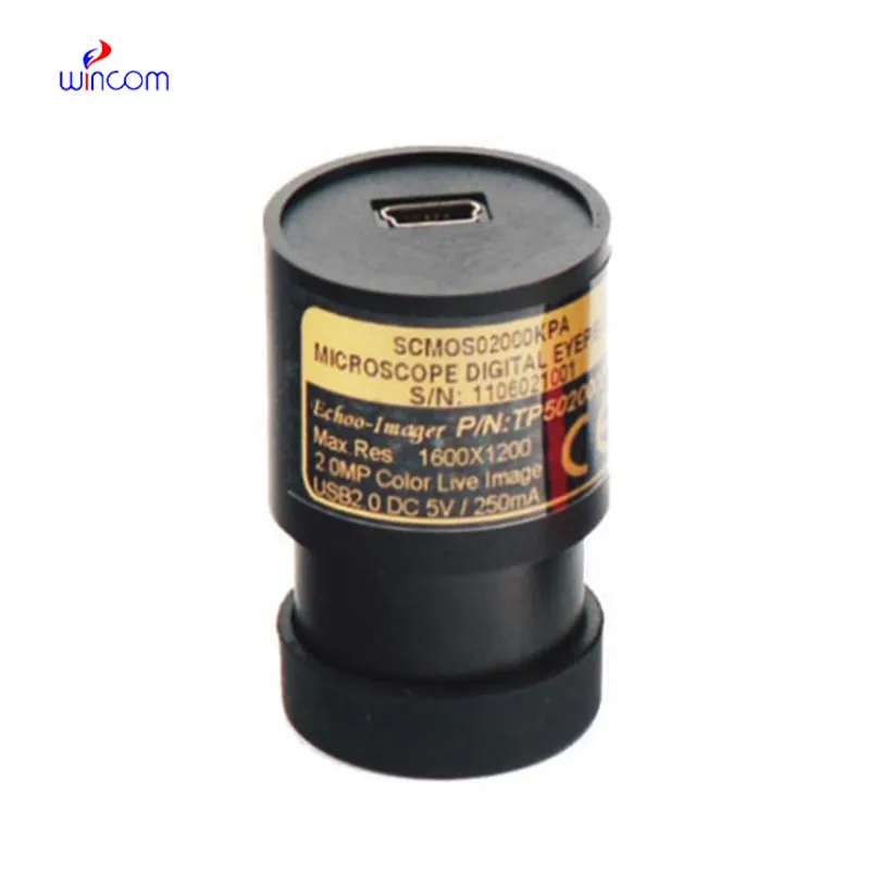
I’ve used several microscopes before, but this one stands out for its sturdy design and smooth magnification control.
To protect the privacy of our buyers, only public service email domains like Gmail, Yahoo, and MSN will be displayed. Additionally, only a limited portion of the inquiry content will be shown.
Could you please provide more information about your microscope range? I’d like to know the magnif...
Hello, I’m interested in your centrifuge models for laboratory use. Could you please send me more ...
E-mail: [email protected]
Tel: +86-731-84176622
+86-731-84136655
Address: Rm.1507,Xinsancheng Plaza. No.58, Renmin Road(E),Changsha,Hunan,China

 af
af
 es
es
 ar
ar
 tr
tr
 sw
sw
 pt
pt
 th
th
 ur
ur
 bn
bn
 ne
ne
 vi
vi
 km
km
 lo
lo
 de
de
 ru
ru
 fi
fi
 nl
nl
 fa
fa
 fr
fr
 ko
ko