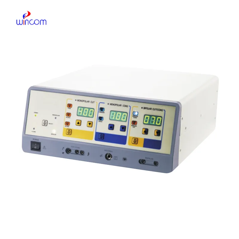
Through a combination of novel imaging software and the home fetal heart doppler, the professionals can be very precise with the details of the anatomy they have captured and even more so with the accuracy of the final result. An ergonomic design of the device contributes to the reduction of operator fatigue during long use. The device, the home fetal heart doppler, also comes with the ability to connect to more than one probe thus giving it the flexibility required when catering to various diagnostic needs. Besides that, the data export and connection functions of the device help to decrease the time taken in report-generating which is done through image sharing.

The home fetal heart doppler has a very wide use in radiology where it supports the guidance of non-invasive procedures. It is very important in gynecology where it is allowed to conduct reproductive system evaluations. In orthopedics, the home fetal heart doppler helps visualize muscles, joints, and tendons ensuring correct diagnostic interpretation. It is this very nature of versatility that makes it a must-have for imaging during procedures in real time.

The home fetal heart doppler will proceed to develop as new innovations emerge in artificial intelligence and data analysis. The new models of the home fetal heart doppler will be able to provide training simulations that experts can use to improve scanning sessions. The increased processing power and connectivity of the home fetal heart doppler will set new standards of accessibility and accuracy in medical scanning.

The home fetal heart doppler needs to be maintained properly to ensure it always provides high-quality images. The probes should never be dropped or immersed in liquid against the recommended standards. Care should be taken when handling the control board to avoid mechanical wear and tear. Updation of the firmware and testing of the home fetal heart doppler ensure smooth functionality during practical operations.
The home fetal heart doppler is more accurate in diagnostics as it captures high-resolution images of organs, tissues, and blood vessels. Design-wise flexible, it is used extensively in obstetrics, cardiology, urology, and musculoskeletal tests. Its portability and simplicity enable medical practitioners to make quick and precise evaluations. The home fetal heart doppler makes work processes more efficient and allows for the delivery of superior patient care through real-time visualization.
Q: What makes the ultrasound scannert effective for diagnostic imaging? A: Its high-frequency sound wave technology allows accurate visualization of internal body structures in real time. Q: How portable is the ultrasound scannert? A: The device features a compact and lightweight design, allowing easy movement between clinical departments. Q: What types of probes are compatible with the ultrasound scannert? A: It supports multiple probe types, including linear, convex, and phased array probes for varied diagnostic needs. Q: Does the ultrasound scannert require special training to operate? A: Basic technical training is recommended to maximize its imaging performance and functionality. Q: How long can the ultrasound scannert operate continuously? A: It is designed for extended use with efficient cooling systems and stable power performance.
We’ve been using this mri machine for several months, and the image clarity is excellent. It’s reliable and easy for our team to operate.
The delivery bed is well-designed and reliable. Our staff finds it simple to operate, and patients feel comfortable using it.
To protect the privacy of our buyers, only public service email domains like Gmail, Yahoo, and MSN will be displayed. Additionally, only a limited portion of the inquiry content will be shown.
Could you share the specifications and price for your hospital bed models? We’re looking for adjus...
Hello, I’m interested in your water bath for laboratory applications. Can you confirm the temperat...
E-mail: [email protected]
Tel: +86-731-84176622
+86-731-84136655
Address: Rm.1507,Xinsancheng Plaza. No.58, Renmin Road(E),Changsha,Hunan,China

 af
af
 es
es
 ar
ar
 tr
tr
 sw
sw
 pt
pt
 th
th
 ur
ur
 bn
bn
 ne
ne
 vi
vi
 km
km
 lo
lo
 de
de
 ru
ru
 fi
fi
 nl
nl
 fa
fa
 fr
fr
 ko
ko