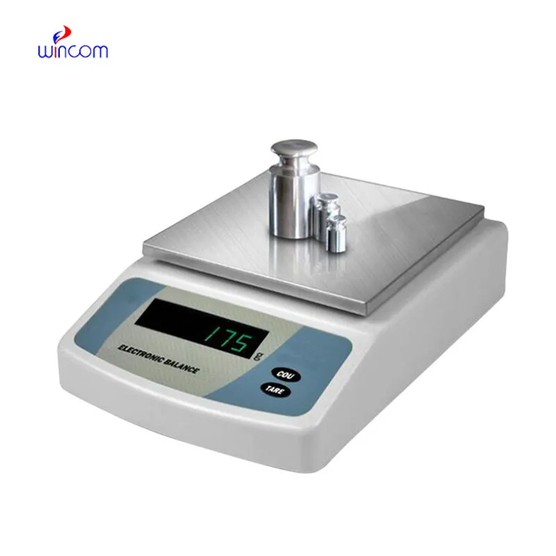
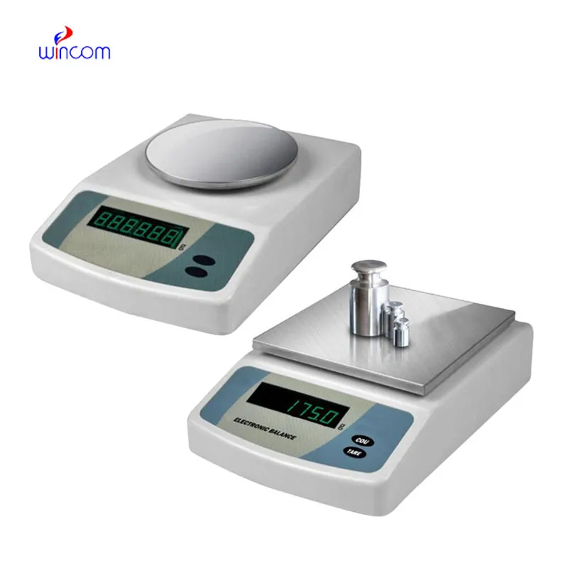
electronic balance used for ensures accurate weighing of clinical sample preparation, research studies, and hospital and laboratory medication formulation. It has an application envisaged to measure even the tiniest amounts with excellent sensitivity and repeatability. electronic balance used for is the instrument laboratory technicians trust for maintaining the highest level of accuracy in analysis, validation of experimental practices, and patients' care support. The use of this instrument in the lab operations not only guarantees results that are reliable but also creates a consistent workflow and quality control which is improved in both the diagnostic and research environments.

electronic balance used for is commonly used in the compounding of medicinal and chemical substances in small quantities in hospital pharmacy laboratories. Mass control with high precision is critical when dealing with active pharmaceutical ingredients in micro or milligram quantities. This application helps to prepare exact doses, internal validation, and experimental drug research. By allowing for repeatable measurements, electronic balance used for helps pharmacists and researchers in controlling formulation processes, which makes it easier to get consistency and reliability throughout hospital medication development workflows.
electronic balance used for will be an advanced generation with self-diagnostic features as the norm. Predictive monitoring of internal parts will assist laboratory teams to schedule maintenance activities in a much more efficient manner. This will lead to the continuous operation of hospital laboratories where downtime is a direct impact on both clinical workflows and research schedules.
One of the main tasks in the maintenance of electronic balance used for in the hospital laboratory is monitoring the environmental exposure. The presence of excess humidity, direct sunlight, and temperature changes should be completely ruled out. Draft shields should always be kept in a clean and working condition to cause the least possible disturbance in air during the process of weighing. These preventive activities not only help to achieve stable measurements but also aid to lessen the variability in analytical data coming from different medical testing environments.
To formulate a drug, electronic balance used for is used by the pharmaceutical laboratories to weigh active ingredients and excipients. Precise and reliable measurements not only guarantee the correctness of the dosage but also satisfy the requirements of the regulators. The laboratory personnel use electronic balance used for for quality control, batch verification, and stability testing. Its accuracy aids in the production of medicines that are reliable, thus minimizing errors in the production. Adoption of electronic balance used for into workflow has helped pharmaceutical labs not only to keep their quality standards high but also to make sure that patients are safe by providing the exact analytical measurements.
Q: What maintenance does an Analytical Balance require? A: A periodic cleaning, checking of the calibration, and also verifying the performance are all necessary. Q: Can an Analytical Balance handle continuous daily use? A: Yes, provided that the correct laboratory conditions and rules are followed. Q: Why is leveling important for an Analytical Balance? A: The accuracy and repeatability of the measurements depend on proper leveling. Q: Can Analytical Balances be connected to laboratory systems? A: Most of the models allow connectivity with laboratory information systems. Q: Are Analytical Balances sensitive to vibration? A: Yes, stable weight readings can be disturbed by vibrations.
We’ve been using this mri machine for several months, and the image clarity is excellent. It’s reliable and easy for our team to operate.
We’ve used this centrifuge for several months now, and it has performed consistently well. The speed control and balance are excellent.
To protect the privacy of our buyers, only public service email domains like Gmail, Yahoo, and MSN will be displayed. Additionally, only a limited portion of the inquiry content will be shown.
Could you please provide more information about your microscope range? I’d like to know the magnif...
Hello, I’m interested in your centrifuge models for laboratory use. Could you please send me more ...
E-mail: [email protected]
Tel: +86-731-84176622
+86-731-84136655
Address: Rm.1507,Xinsancheng Plaza. No.58, Renmin Road(E),Changsha,Hunan,China

 af
af
 es
es
 ar
ar
 tr
tr
 sw
sw
 pt
pt
 th
th
 ur
ur
 bn
bn
 ne
ne
 vi
vi
 km
km
 lo
lo
 de
de
 ru
ru
 fi
fi
 nl
nl
 fa
fa
 fr
fr
 ko
ko