With the cutting-edge imaging processors, the b-scanner ultrasound facilitates real-time, high-resolution images that are paramount in the detection of subtle physiological changes by clinicians. The display is user-friendly for easy parameter modification as well as image marking. The b-scanner ultrasound exhibits the mix of effectiveness, mobility, and reliability for a huge range of diagnostic procedures.

The vast clinical applications of the b-scanner ultrasound technology made it possible for nephrology to monitor kidney function efficiently and detect abnormalities in kidney structure. In the frontiers of endocrinology, the obtained data can reveal even the smallest nodules in the glands. The b-scanner ultrasound is also a surgical device for blood flow patterns and vessel integrity.

The b-scanner ultrasound should integrate with intelligent diagnostic ecosystems and communicate effortlessly with smartphones and electronic records. The synchronized exchange of data in real-time should enable constant patient observation. The next version should focus on improved design, better processing power of artificial intelligence algorithms, and enhanced reconstruction functions.

In order to retain the accuracy of the b-scanner ultrasound, it is important for operators to check the cables and connections of the transducers for evidence of wear. After each use, the surfaces should be wiped clean using non-abrasive cleaners. The b-scanner ultrasound should be turned off properly and covered to prevent dust from collecting. Regular checks by trained personnel should be done.
The b-scanner ultrasound is a critical diagnostic tool used across all the medical specialties for imaging internal organs, tissues, and blood flow in real time. It generates high-resolution images with no patient radiation exposure using high-frequency sound waves. The b-scanner ultrasound ensures precise monitoring across obstetrics, cardiology, and emergency medicine. Its portability and simplicity ensure it is useful in both field and clinical settings, enhancing diagnostic efficiency and precision.
Q: What is the primary function of an ultrasound scannert? A: Ultrasound scanners are designed to create real-time images of internal organs, tissues, and blood flow using high-frequency sound waves. Q: How does the ultrasound scannert ensure clear imaging results? A:It uses advanced converter technology and digital processing to enhance image clarity and contrast. Q: In what medical fields is the ultrasound scannert commonly used? A: It is widely used in obstetrics, cardiology, urology, radiology, and emergency medicine. Q: Is the ultrasound scannert safe for repeated use? A: Yes, it is non-invasive and does not emit radiation, making it safe for frequent diagnostic applications. Q: Can the ultrasound scannert store and share imaging data? A: Yes, it supports data storage, retrieval, and digital transfer for easy integration with hospital systems.
The microscope delivers incredibly sharp images and precise focusing. It’s perfect for both professional lab work and educational use.
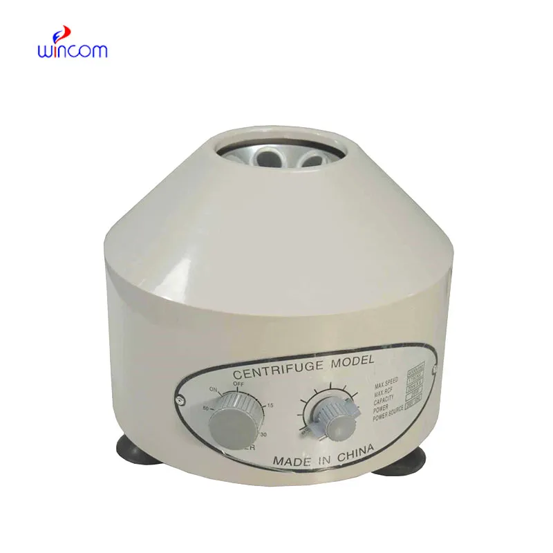
The centrifuge operates quietly and efficiently. It’s compact but surprisingly powerful, making it perfect for daily lab use.
To protect the privacy of our buyers, only public service email domains like Gmail, Yahoo, and MSN will be displayed. Additionally, only a limited portion of the inquiry content will be shown.
We’re currently sourcing an ultrasound scanner for hospital use. Please send product specification...
We are planning to upgrade our imaging department and would like more information on your mri machin...
E-mail: [email protected]
Tel: +86-731-84176622
+86-731-84136655
Address: Rm.1507,Xinsancheng Plaza. No.58, Renmin Road(E),Changsha,Hunan,China

 af
af
 es
es
 ar
ar
 tr
tr
 sw
sw
 pt
pt
 th
th
 ur
ur
 bn
bn
 ne
ne
 vi
vi
 km
km
 lo
lo
 de
de
 ru
ru
 fi
fi
 nl
nl
 fa
fa
 fr
fr
 ko
ko